Resolution and Electronic Imaging
- Resolvable Detail
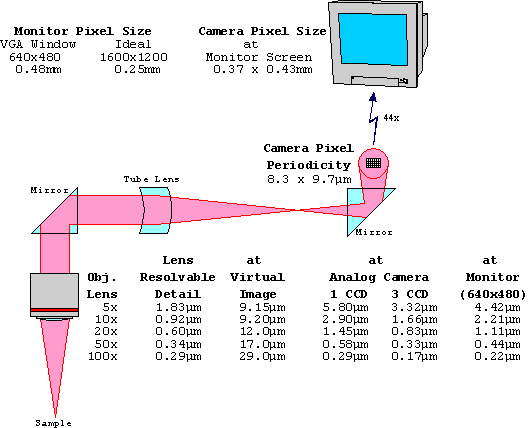
- The smallest resolvable detail imaged by an objective lens can be
estimated by 0.550µm/2xNumerical Aperture. That detail appears in
the virtual image produced by the objective lens, enlarged by the
magnification of the lens. The detail in the virtual image is
projected via a tube lens onto the surface of a CCD chip in a color
camera. The smallest resolvable detail in the virtual image
observable with the camera is determined by the pixel periodicity of
the CCD chip. The output of the camera goes through a video
controller board for pixel interpolation and is transferred to the
monitor for display. The smallest resolvable detail observable at
the monitor is determined by the monitor pixel periodicity which is
usually based on the display mode. In very high resolution systems
observable resolution is limited by the Red-Green-Blue triad
periodicity of the cathode ray tube.
-
- The diagram below shows an example of the smallest resolvable
detail at each level in an imaging path from an objective lens to a
monitor. The resolution-limiting component is the one that has the
largest detail size shown for that objective lens.
- Field-of-View
-
The field-of-view for a microscope with oculars is
equal to the Ocular Field # / Objective Lens Magnification (if there
are no magnifying intermediate lenses). For Field # 20 oculars, the
field-of-view is shown below, along with the area displayed on a
typical electronic imaging system.
| Obj |
Field-of-View
with Oculars |
Video |
| Lens |
Total
Diameter |
Perceived
Area |
Displayed
Area |
| 5x |
4.0
mm |
2.40
x 2.00 mm |
1.20
x 0.90 mm |
| 10x |
2.0
mm |
1.20
x 1.00 mm |
0.60
x 0.45 mm |
| 20x |
1.0
mm |
0.60
x 0.50 mm |
0.30
x 0.22 mm |
| 50x |
0.4
mm |
0.24
x 0.20 mm |
0.12
x 0.09 mm |
| 100x |
0.2
mm |
0.12
x 0.10 mm |
0.06
x 0.045mm |
|
- When performing rapid screening through oculars, the area that
information is gleaned from is not the total area illuminated1,2.
The perceived area shown in the table is an average (some see more,
while others see less). But the reality is that the best one can
expect to glean information from is 75% of the diameter on the
horizontal axis and 66% on the vertical axis. The lower limit is
50%/33%. The Perceived Area data reflects the average. Since a video
image is 100% perceived, to achieve the same screening area, a 2x
lower objective is required.
|

Total Diameter/Perceived Area |
- "How Did I Miss Those Cells?" Gary W. Gill, CT (ASCP), ADVANCE
for Medical Laboratory Professionals, Nov. 20, 1995
- "Visual Conspicuity, visual search and fixation tendencies of
the eye" Engel F.L., Vision Res. 17:95-108, 1977
|


